详细说明
Product Brief
Product Overview
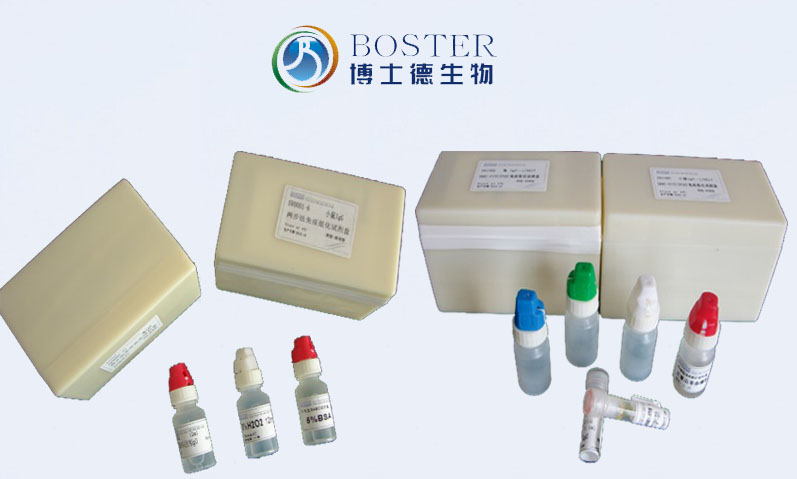
Product Name Goat Anti-Rabbit IgG(H+L) Secondary Antibody, 10 nm Colloidal Gold Conjugate Synonyms 10 nm Colloidal Gold Labeled Secondary Antibody, Goat Anti-Rabbit IgG; 10 nm Colloidal Gold Conjugated Goat Anti-Rabbit IgG; Goat Anti-Rabbit IgG 10 nm Colloidal Gold Secondary Antibody Description Goat Anti-rabbit IgG secondary antibody, 10 nm Colloidal Gold Conjugate, for detection, localization, distribution and quantification of target proteins at an ultrastructural level via indirect immunogold staining in IHC-P, IHC-F, ICC, or EM. Reagent Type Colloidal gold conjugated secondary antibody Conjugate Colloidal gold, 10 nm Host Goat Target Species Rabbit Antibody Class IgG Clonality Polyclonal Immunogen Whole molecule rabbit IgG Purification Immunoaffinity chromatography, solid phase adsorbed with human serum proteins Specificity Rabbit IgG specific: no cross-reactivity with mouse/bovine IgG Form Supplied Liquid: concentrated buffered stock solution Formulation 0.5 mg colloidal gold-conjugated secondary antibody
0.01 M PBS (PH 7.4)
0.01% Thimerosal
50% glycerolPack Size 0.5ml/1ml
Concentration 0.1 mg/ml Application Electron Microscopy, IHC-P, IHC-F, ICC Storage 4C for 1 year Precautions FOR RESEARCH USE ONLY. NOT FOR DIAGNOSTIC OR CLINICAL USE Assay Information
Sample Type Goat primary-antibody-probed ultrathin sliced formalin-fixed paraffin-embedded (FFPE) tissue sections(IHC-P), ultrathin thawed frozen sections(IHC-F), cultured cells Assay Type Immunoanalytical Technique Indirect immunodetection of target protein via reporter-labeled biotin-binding detection systems Assay Purpose Protein detection/quantification Equipment Needed Light microscope, scanning electron microscope or transmission electron microscope, micrograph or scan Main Advantages
Specific High signal-to-noise ratio High Signal Amplification Multiple secondary antibodies can bind to a single primary antibody; label size is larger than tissue structures; much more intense stain than conventional HRP and PAP techniques; additional silver enhancement can be applied Fast Fewer processing steps - no need to add a substrate; Less optimization required compared to enzymatic detection; Generates strong signals in a relatively short time span; signal can be observed directly Quantifieable The digital nature of the gold signal + high precision in allocating gold labels to defined structures makes it easy to count and quantify Easy to Use Supplied in a workable liquid format Flexible Diversity of non-overlapping particle sizes for labeling at different working magnifications, suitable for single or multi-label detection, may be used as a probe in immunoblotting, light microscopy, fluorescence microscopy or electron microscopy, ability to label sections from the same block for both light and electron microscopy, can be used to localize certain antigens in sectioned tissue or cultured cells, excellent labelling in wax, resin embedded or frozen sections, a variety of tissue stains can be used to counterstain the dense black reaction product after silver enhancement Stability Gold particles bind proteins rapidly and stably - permanent label, no fading, macromolecule localization and distribution can be recorded using specialized photography Background
Most commonly, secondary antibodies are generated by immunizing the host animal with a pooled population of immunoglobulins from the target species. The host antiserum is then purified through immunoaffinity chromatography to remove all host serum proteins, except the specific antibody of interest, and then modified with antibody fragmentation, label conjugation, etc., to generate highly specific detection reagents. Secondary antibodies can be conjugated to a large number of labels, including enzymes, biotin, fluorescent dyes/proteins, or gold particles. Here, the antibody provides the specificity to locate the protein of interest by recognizing a primary antibody that targets a particular antigen, and the label generates a detectable signal in the area of the formed immune complexes. The label of choice depends upon the experimental application.
Immunolabeling with gold particles—also known as immunogold labeling, immunogold staining, or immunocolloidal gold technique—is a technique that uses colloidal gold as a marker. Colloidal gold is a negatively charged hydrophobic suspension, formed by electron dense metallic particles which have large specific surface area. It binds to macromolecules (IgG, Protein A or G) rapidly and stably by noncovalent electrostatic adsorption, preserving their natural biological activity. It has been used as a marker particle for the past 2 decades because of its simplicity of preparation, high resolution, good biocompatibility and tissue penetration and hence its suitability for ultrastructural studies and quantitative immunostaining. Due to variability in particle sizes and its high electron density, immunocolloidal gold technique is suitable for single or multi-label detection under immune-electron microscopes as well as light microscopes. It is used regularly with scanning electron microscopy (SEM) and transmission electron microscopy (TEM) to successfully identify the areas within cells and tissues where antigens are located. The high resolution of the technique allows to study structure–function relationships in the microenvironment of cells and tissues, and also explore protein distribution in cellular and extracellular components. Smaller gold particles are found to produce higher labeling intensity of the target antigen because of reduced steric hindrance. However, if steric hindrance does not make a significant difference, then larger gold particles could produce stronger color intensity under the light microscope. When smaller gold particles (1-5 nm) are used, the visualization is usually improved using silver-enhancement methods. Gold can also be attached to protein A or protein G instead of a secondary antibody, as these proteins bind mammalian IgG Fc regions in a non-specific way.
Instructions
胶体金稀释液:PBS 或 TBS 缓冲液,加入试剂级 BSA 至 1%。 使用上述胶体金稀释液,免疫电镜按 1:20—50 稀释,免疫组化按 1:50—100 稀释。胶体金 稀释后溶液应于当天使用,未用完的应弃置。


