详细说明
概述
产品名称Anti-Bmi1抗体[EPR3745(2)]
描述
兔单克隆抗体[EPR3745(2)] to Bmi1
经测试应用WB,IHC-P,ICC/IF
种属反应性
与反应: Rat, Human
免疫原
A synthetic peptide, corresponding to residues in Human Bmi1 (UniProt P35226).
阳性对照
WB: K562, SAOS-2, SW480, MOLT4, PC-12 and HT1080 cell lysates. IHC-P: Human tonsil, colonic adenocarcinoma, lung adenocarcinoma, breast carcinoma and thyroid gland carcinoma tissues. ICC/IF: SW480 and HeLa cells.
常规说明
This product is a recombinant rabbit monoclonal antibody.
We are constantly working hard to ensure we provide our customers with best in class antibodies. As a result of this work we are pleased to now offer this antibody in purified format. We are in the process of updating our datasheets. The purified format is designated ‘PUR’ on our product labels. If you have any questions regarding this update, please contact our Scientific Support team.
Produced using Abcam’s RabMAb® technology. RabMAb® technology is covered by the following U.S. Patents, No. 5,675,063 and/or 7,429,487.
Mouse: Internal data indicated that the antibody is not suitable for WB application in mouse species.
Alternative versions available:
性能
形式Liquid
存放说明Shipped at 4°C. Store at +4°C short term (1-2 weeks). Upon delivery aliquot. Store at -20°C. Stable for 12 months at -20°C.
存储溶液pH: 7.20
Preservative: 0.01% Sodium azide
Constituents: 59% PBS, 40% Glycerol, 0.05% BSA纯度Protein A purified
克隆单克隆
克隆编号EPR3745(2)
同种型IgG
研究领域
Cancer
Oncoproteins/suppressors
Oncoproteins
Other
Cancer
Cell cycle
Cell differentiation
Stem Cells
Hematopoietic Progenitors
Intracellular Molecules
Epigenetics and Nuclear Signaling
Transcription
Cancer susceptibility
Proto-oncogenes
Epigenetics and Nuclear Signaling
Chromatin Remodeling
Polycomb Silencing
PRC2
Cell Biology
Cell Cycle
Cell differentiation
Anti-Bmi1 antibody [EPR3745(2)] 图像
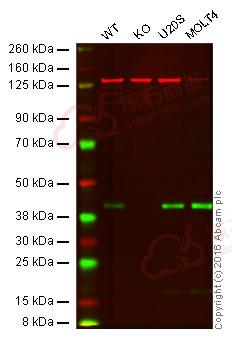
![Western blot - Anti-Bmi1 antibody [EPR3745(2)] (ab126783)](http://img.lianshimall.com/statics/attachment/goods/pl20160426/abcamMainImgPrimary/detail/ab12/ab126783OWB.jpg)
Western blot - Anti-Bmi1 antibody [EPR3745(2)] (ab126783)
Predicted band size : 36 kDa
Lane 1: Wild-type HAP1 cell lysate (20 µg)
Lane 2: Bmi1 knockout HAP1 cell lysate (20 µg)
Lane 3: U2OS cell lysate (20 µg)
Lane 4: Molt-4 cell lysate (20 µg)
Lanes 1 - 4: Merged signal (red and green). Green - ab126783 observed at 42 kDa. Red - loading control, ab8245, observed at 37 kDa.
ab126783 was shown to specifically react with Bmi1 when Bmi1 knockout samples were used. Wild-type and Bmi1 knockout samples were subjected to SDS-PAGE. ab126783 and ab8245 (loading control to GAPDH) were both diluted at 1/10 000 and incubated overnight at 4°C. Blots were developed with goat anti-rabbit IgG (H + L) and goat anti-mouse IgG (H + L) secondary antibodies at 1/10 000 dilution for 1 h at room temperature before imaging.![Immunohistochemistry (Formalin/PFA-fixed paraffin-embedded sections) - Anti-Bmi1 antibody [EPR3745(2)] (ab126783)](http://img.lianshimall.com/statics/attachment/goods/pl20160426/abcamMainImgPrimary/detail/ab12/ab126783HC1.jpg)
Immunohistochemistry (Formalin/PFA-fixed paraffin-embedded sections) - Anti-Bmi1 antibody [EPR3745(2)] (ab126783)
Immunohistochemistry (Formalin/PFA-fixed paraffin-embedded sections) analysis of human breast carcinoma tissue labelling Bmi1 with purified ab126783 at 1/500. Heat mediated antigen retrieval was performed using Tris/EDTA buffer pH 9. ab97051, a HRP-conjugated goat anti-rabbit IgG (H+L) was used as the secondary antibody (1/500). Negative control using PBS instead of primary antibody. Counterstained with hematoxylin.
![Immunocytochemistry/ Immunofluorescence - Anti-Bmi1 antibody [EPR3745(2)] (ab126783)](http://img.lianshimall.com/statics/attachment/goods/pl20160426/abcamMainImgPrimary/detail/ab12/ab126783IF1.jpg)
Immunocytochemistry/ Immunofluorescence - Anti-Bmi1 antibody [EPR3745(2)] (ab126783)
Immunocytochemistry/Immunofluorescence analysis of HeLa cells labelling Bmi1 with purified ab126783 at 1/500. Cells were fixed with 4% paraformaldehyde and permeabilized with 0.1% Triton X-100. ab150077, an Alexa Fluor® 488-conjugated goat anti-rabbit IgG (1/500) was used as the secondary antibody. DAPI (blue) was used as the nuclear counterstain. ab7291, a mouse anti-tubulin (1/1000) and ab150120, an Alexa Fluor® 594-conjugated goat anti-mouse IgG (1/500) were also used.
Control 1: primary antibody (1/500) and secondary antibody, ab150120, an Alexa Fluor® 594-conjugated goat anti-mouse IgG (1/500).
Control 2: ab7291 (1/1000) and secondary antibody, ab150077, an Alexa Fluor® 488-conjugated goat anti-rabbit IgG (1/500).
![Western blot - Anti-Bmi1 antibody [EPR3745(2)] (ab126783)](http://img.lianshimall.com/statics/attachment/goods/pl20160426/abcamMainImgPrimary/detail/ab12/ab126783WB2.jpg)
Western blot - Anti-Bmi1 antibody [EPR3745(2)] (ab126783)
Anti-Bmi1 antibody [EPR3745(2)] (ab126783) at 1/20000 dilution (purified) + PC-12 cell lysate at 20 µg
Secondary
Peroxidase-conjugated goat anti-rabbit IgG, (H+L) at 1/1000 dilution
Predicted band size : 36 kDa
Observed band size : 40 kDa (why is the actual band size different from the predicted?)
Blocking and dilution buffer: 5% NFDM/TBST.
![Western blot - Anti-Bmi1 antibody [EPR3745(2)] (ab126783)](http://img.lianshimall.com/statics/attachment/goods/pl20160426/abcamMainImgPrimary/detail/ab12/ab126783WB1.jpg)
Western blot - Anti-Bmi1 antibody [EPR3745(2)] (ab126783)
All lanes : Anti-Bmi1 antibody [EPR3745(2)] (ab126783) at 1/20000 dilution (purified)
Lane 1 : K562 cell lysate
Lane 2 : SAOS-2 cell lysate
Lane 3 : SW480 cell lysate
Lane 4 : Molt-4 cell lysate
Lysates/proteins at 20 µg per lane.
Secondary
Peroxidase-conjugated goat anti-rabbit IgG, (H+L) at 1/1000 dilution
Predicted band size : 36 kDa
Observed band size : 40 kDa (why is the actual band size different from the predicted?)
Blocking and dilution buffer: 5% NFDM/TBST.
Western blot - Anti-Bmi1 antibody [EPR3745(2)] (ab126783)
All lanes : Anti-Bmi1 antibody [EPR3745(2)] (ab126783) at 1/10000 dilution (unpurified)
Lane 1 : K562 cell lysate
Lane 2 : SAOS-2 cell lysate
Lane 3 : SW480 cell lysate
Lane 4 : MOLT4 cell lysate
Lane 5 : HT1080 cell lysate
Lysates/proteins at 10 µg per lane.
Secondary
HRP-conjugated goat anti-rabbit IgG at 1/2000 dilution
Predicted band size : 36 kDa
![Immunohistochemistry (Formalin/PFA-fixed paraffin-embedded sections) - Anti-Bmi1 antibody [EPR3745(2)] (ab126783)](http://img.lianshimall.com/statics/attachment/goods/pl20160426/abcamMainImgPrimary/detail/ab12/ab1267833-4.jpg)
Immunohistochemistry (Formalin/PFA-fixed paraffin-embedded sections) - Anti-Bmi1 antibody [EPR3745(2)] (ab126783)
Immunohistochemistry (Formalin/PFA-fixed paraffin-embedded sections) analysis of human normal tonsil tissue labelling Bmi1 with unpurifiied ab126783.
![Immunohistochemistry (Formalin/PFA-fixed paraffin-embedded sections) - Anti-Bmi1 antibody [EPR3745(2)] (ab126783)](http://img.lianshimall.com/statics/attachment/goods/pl20160426/abcamMainImgPrimary/detail/ab12/ab1267833-5.jpg)
Immunohistochemistry (Formalin/PFA-fixed paraffin-embedded sections) - Anti-Bmi1 antibody [EPR3745(2)] (ab126783)
Immunohistochemistry (Formalin/PFA-fixed paraffin-embedded sections) analysis of human colonic adenocarcinoma tissue labelling Bmi1 with unpurifiied ab126783.
![Immunohistochemistry (Formalin/PFA-fixed paraffin-embedded sections) - Anti-Bmi1 antibody [EPR3745(2)] (ab126783)](http://img.lianshimall.com/statics/attachment/goods/pl20160426/abcamMainImgPrimary/detail/ab12/ab1267833-6.jpg)
Immunohistochemistry (Formalin/PFA-fixed paraffin-embedded sections) - Anti-Bmi1 antibody [EPR3745(2)] (ab126783)
Immunohistochemistry (Formalin/PFA-fixed paraffin-embedded sections) analysis of human lung adenocarcinoma tissue labelling Bmi1 with unpurifiied ab126783.
![Immunohistochemistry (Formalin/PFA-fixed paraffin-embedded sections) - Anti-Bmi1 antibody [EPR3745(2)] (ab126783)](http://img.lianshimall.com/statics/attachment/goods/pl20160426/abcamMainImgPrimary/detail/ab12/ab1267833-7.jpg)
Immunohistochemistry (Formalin/PFA-fixed paraffin-embedded sections) - Anti-Bmi1 antibody [EPR3745(2)] (ab126783)
Immunohistochemistry (Formalin/PFA-fixed paraffin-embedded sections) analysis of human thyroid gland carcinoma tissue labelling Bmi1 with unpurifiied ab126783.
Immunocytochemistry/ Immunofluorescence - Anti-Bmi1 antibody [EPR3745(2)] (ab126783)
Immunocytochemistry/Immunofluorescence analysis of SW480 cells labelling Bmi1 with unpurified ab126783 at a dilution of 1/100.


![Anti-Blood Group Antigen Precursor antibody [EPR6205] 40µl](https://yunshiji.oss-cn-shenzhen.aliyuncs.com/202407/25/nbgdgpchhpd.jpg)
![Anti-Blood Group Antigen Precursor antibody [EPR6205] 100µl](https://yunshiji.oss-cn-shenzhen.aliyuncs.com/202407/25/w35rxf2jhgg.jpg)
![Anti-BLNK antibody [Y491] 10µl](https://yunshiji.oss-cn-shenzhen.aliyuncs.com/202407/25/ewoke0zmvuo.jpg)
![Anti-BLNK antibody [Y491] 40µl](https://yunshiji.oss-cn-shenzhen.aliyuncs.com/202407/25/jk5zkwrn3uy.jpg)
![Anti-BLNK antibody [Y491] 100µl](https://yunshiji.oss-cn-shenzhen.aliyuncs.com/202407/25/dkqjvkexwbl.jpg)
![Western blot - Anti-Bmi1 antibody [EPR3745(2)] (ab126783)](http://img.lianshimall.com/statics/attachment/goods/pl20160426/abcamMainImgPrimary/detail/ab12/ab1267833-1.JPG)
![Immunocytochemistry/ Immunofluorescence - Anti-Bmi1 antibody [EPR3745(2)] (ab126783)](http://img.lianshimall.com/statics/attachment/goods/pl20160426/abcamMainImgPrimary/detail/ab12/ab1267833-3.JPG)


 粤公网安备44196802000105号